38 label the transmission electron microscope image of a chloroplast below
41 label the transmission electron microscope image of a ... (A, B) Ultrastructure of chloroplasts and mitochondria... DP Biology: Ultrastructure of cells quiz 1.2 The electron microscope image below shows an organelle ... Amazing 27 Things Under The Microscope With Diagrams Amazing 27 Things Under The Microscope With Diagrams February 20, 2022 by Anupama Sapkota Note: Each image source is given below in this post of respective subheadings. Table of Contents 1. Amoeba under the microscope Direct observation Observation after staining 2. Algae under the microscope Chlorophyta Chromophyta Cryptophyta Rhodophyta
DP Biology (Cells: 1.1, 1.2, 1.5, 6.3) Quiz - Quizizz The image shows a phagocytic white blood cell as seen with a transmission electron microscope. Which features can be found both within this cell and in a photosynthetic bacterium? answer choices chloroplasts multiple nuclei 70s ribosomes lysosomes Question 2 30 seconds Q. The image shows an electron micrograph of a fungus, Candida albicans.

Label the transmission electron microscope image of a chloroplast below
Cells and magnification Flashcards - Quizlet 6) a- Figure 2 shows a photos of part of a mitochondrion from a mouse liver cell taken using a transmission electron microscope at x62800 magnification Produce a scientific drawing of the mitochondria in figure 2 Label the following part of the mitochondrion on your drawing -Matrix-Crista (4 marks) Labeling the Cell Flashcards | Quizlet Label the transmission electron micrograph of the nucleus. membrane bound organelles golgi apparatus, mitochondrion, lysosome, peroxisome, rough endoplasmic reticulum nonmembrane bound organelles ribosomes, centrosome, proteasomes cytoskeleton includes microfilaments, intermediate filaments, microtubules Identify the highlighted structures Examining epithelial tissue under the microscope - Course Hero There are three basic shapes used to classify epithelial cells. A squamous epithelial cell looks flat under a microscope. A cuboidal epithelial cell looks close to a square. A columnar epithelial cell looks like a column or a tall rectangle. A few epithelial layers are constructed from cells that are said to have a transitional shape.
Label the transmission electron microscope image of a chloroplast below. Cell and Molecular Biology part 1 Flashcards - Cram.com Please select the correct language below. Front audio not yet available for this language ... Mannose-6-phosphate is a molecular address label which directs protein to the lysosome. Which of the followinf enzymes would be labled with mannose-6-phosphate? ... Transmission electron microscope images of bacteria show that they also have a nucleus ... PDF General Biology 1 Lab #4: Cells - Brazosport College 5. Observe the specimen under the microscope. Remember: First find water plant cells using 4X objective, then change to 10X and focus and draw, then turn to 40X and draw. 6. Draw a few water plant cells below, labeling the nucleus, cytoplasm, cell wall, and the chloroplasts. 7. Leave under the microscope for 20 minutes then check to see if you ... Quia - AP Chapter 6 - Cells (basic) What type of electron microscope produced this image?, transmission electron microscope (TEM), The cell below is a(n) _____ cell., animal, The cell below is a(n) _____ cell., plant, The ____ is the simplest collection of matter that can live. cell: The first microscopes, as well as the microscopes that we use in lab, are called ____. light ... Solved Part 1 (1 point) In the electron micrograph, identify - Chegg Part 1 (1 point) In the electron micrograph, identify the bacterial cell features by dragging the labels to their targets. Cell wall D A Plasma membrane Cytoplasm Outer membrane B с 1 um Q Courtesy of E. Kellenberger What is a drawback to using light microscopy? Choose one: o O A. It cannot be used to view structures smaller than a bacterium. B.
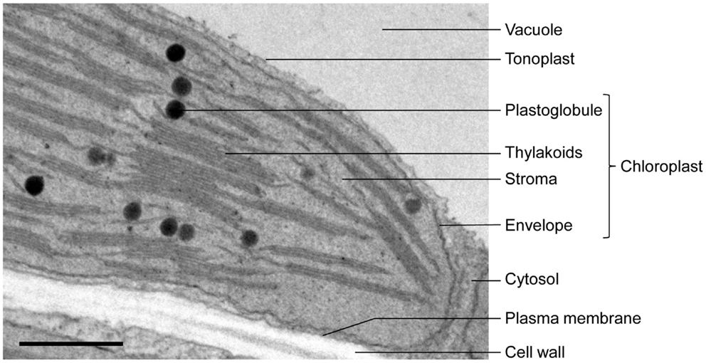
Transmission Electron Microscope (With Diagram) Finally, the electrons are focused by an electromagnetic projector lens (instead of an ocular lens as in a light microscope) on a screen or photographic plate. The final image in a TEM is known as transmission electron micrograph. The salts of some heavy metals, e.g., lead; osmium, tungsten and uranium are often used for staining. Classical transmission electron microscopy (TEM) led to ... - SpringerLink Thin section electron micrograph of a chemically fixed chloroplast in a young tobacco leaf. The chloroplast lies flat against the plasma membrane and the cell wall (CW) and presents a more or less elliptical outline. The stacked grana thylakoids (GT) are interconnected by non-stacked stroma thylakoids (ST). Instruments of Microscopy | Microbiology | | Course Hero Differential interference contrast (DIC) microscopes (also known as Nomarski optics) are similar to phase-contrast microscopes in that they use interference patterns to enhance contrast between different features of a specimen. In a DIC microscope, two beams of light are created in which the direction of wave movement (polarization) differs. (PDF) Field-emission scanning electron microscopy of the ... - Academia.edu specimens were incubated in 1/200 c4 antiactin figure 1 shows a typical image of a fractured higher- for 30 min followed, after washing in pbs, by 1/25 goat anti-mouse igg-10 nm diameter gold (amersham) for 30 min, washed in pbs, plant cell obtained by the osmium maceration proce- postfixed in 0.5% oso4 in pbs for 5 min at 4 ~ washed in dhzo, d …
PDF Chloroplasts Structure and Function Factsheet The biconvex shape of the chloroplast is yet another way of increasing surface area to maximise absorption of light energy Sometimes in the exam you will be presented with an electron micrograph of a chloroplast. Usually, the first question simply asks you to label it. Typical Exam Question Label parts A B & C A B C Answer A - stroma; Looking at the Structure of Cells in the Microscope Images of Surfaces Can Be Obtained by Scanning Electron Microscopy. A scanning electron microscope (SEM) directly produces an image of the three-dimensional structure of the surface of a specimen. The SEM is usually a smaller, simpler, and cheaper device than a transmission electron microscope. chloroplast | Definition, Function, Structure, Location, & ... chloroplast, structure within the cells of plants and green algae that is ... False-colour transmission electron micrograph of a chloroplast in a bean leaf. Chlorophyll catabolism precedes changes in chloroplast structure and ... Ultrathin sections (70-90 nm) were viewed and photographed with a FEI Tecnai SPIRIT (FEI, Eidhoven, Netherlands) transmission electron microscope operating at 120 kV and equipped with an EAGLE CCD Camera. Measurements of chloroplasts, starch bodies, and plastoglobules were carried out using Fiji (ImageJ). 2.8 Mass spectrometry

31 Label The Transmission Electron Microscope Image Of A Chloroplast ...
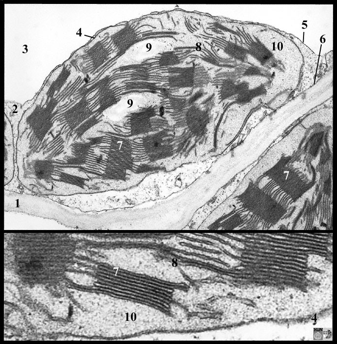
Electron Micrographs of Cell Organelles - Biology Discussion This is an electron-micrograph of plastid or chloroplast, which is an integral component of all green plant leaves and is characterized by following features (Fig. 15 & 16): (1) They may be spheroidal, ovoid, stellate or collar shaped and differ in size and number in different cells.
Basisanatomie
AP Biology Describe the principles, advantages, and limitations of the light microscope, transmission electron microscope, and scanning electron microscope.

32 Label The Transmission Electron Microscope Image Of A Chloroplast ...
The Transmission Electron Microscope | CCBER Transmission electron microscopes (TEM) are microscopes that use a particle beam of electrons to visualize specimens and generate a highly-magnified image. TEMs can magnify objects up to 2 million times. In order to get a better idea of just how small that is, think of how small a cell is.
Frontiers | When Proteomics Reveals Unsuspected Roles: The ...
Electron microscopes - Cell structure - Edexcel - BBC Bitesize the transmission electron microscope (TEM) is used to examine thin slices or sections of cells or tissues the scanning electron microscope (SEM) has a large depth of field so can be used to examine...

BIOLOGY PAPER 1 - KCSE 2019 JOINT PRE MOCK EXAMINATION NAMBALE
Part 3. Structure of The Plant Leaf and Chloroplasts Study this transmission electron micrograph of a spinach leaf cell, locate a chloroplast and capture the image for labeling. The micrograph is displayed as if using a "virtual electron microscope", so you will need to magnify the image and move to a region that contains the clearest view of chloroplast internal structures.
Post a Comment for "38 label the transmission electron microscope image of a chloroplast below"