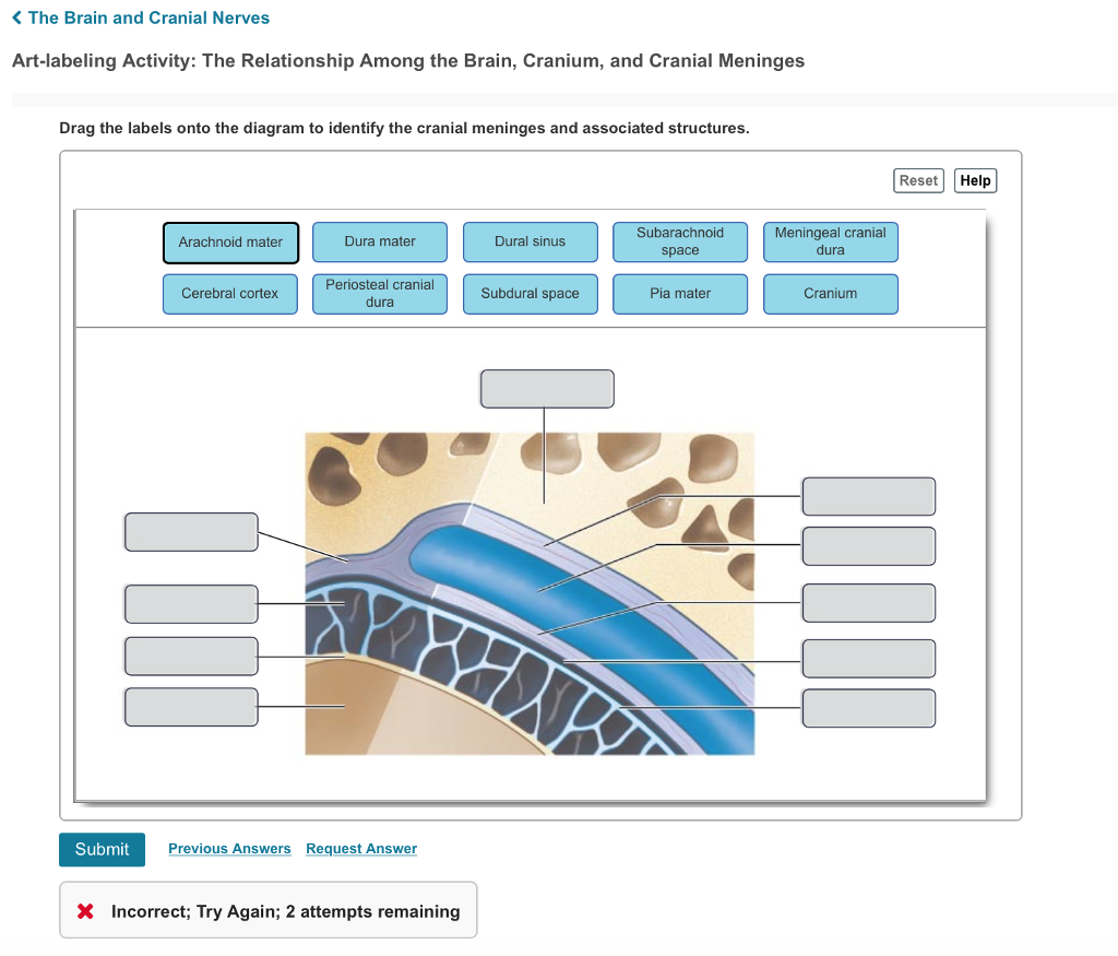
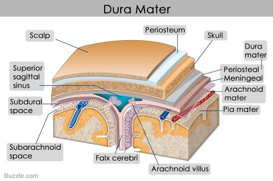
42 correctly label the following meninges and associated structures.
Meninges: Function and Layers, and Health Problems - ThoughtCo The meninges functions primarily to protect and support the central nervous system (CNS). It connects the brain and spinal cord to the skull and spinal canal. The meninges forms a protective barrier that safeguards the sensitive organs of the CNS against trauma. It also contains an ample supply of blood vessels that deliver blood to CNS tissue. Mesentery: Anatomy, functions and clinical points | Kenhub This decreases the friction between the adjacent visceral surfaces and allows some movement of the organs that occur during digestion. The mesentery attaches the intestines to the abdominal wall, and also helps storing the fat and allows the blood and lymph vessels, as well as the nerves, to supply the intestines. Key facts.
From outermost to innermost, what are the names and the ... - Socratic Dura mater Arachnoid mater Pia mater From outermost to innermost, Meninges around the brain has three layers : 1. The dura mater 2. the arachnoid mater and 3. the pia mater. The dura mater has two layers : periosteal and meningeal. There is a space between the dura mater and the arachnoid mater, called subdural space. There is also a space between the arachnoid and the pia mater, which is ...

Correctly label the following meninges and associated structures.
Solved Correctly label the following meninges and associated - Chegg Science. Anatomy and Physiology. Anatomy and Physiology questions and answers. Correctly label the following meninges and associated structures Arachnoid valus Arachnoid maler Meningeal layer of dura Pia mater Subarachnoid space Periosteal layer of dura Fabcccrobni. Label the landmarks of the skull in the figure below Several of these are described on the following pages. Locating Body Landmarks. Anterior Body Landmarks. Identify and use anatomical terms to correctly label the following regions on Figure 1:. i miss you message for her long distance. Bumps and grooves of the brain. In humans, the lobes of the brain are divided by a number of bumps and grooves. What midbrain structure is a visual reflex center Superior colliculi of the tectal plate The Meninges of the Brain Correctly label the following meninges and associated structures. The Flow of Cerebrospinal Fluid Place a single word into each sentence to make it correct. Not all terms will be used.
Correctly label the following meninges and associated structures.. The Meninges - Dura - Arachnoid - Pia - TeachMeAnatomy The meninges refer to the membranous coverings of the brain and spinal cord. There are three layers of meninges, known as the dura mater, arachnoid mater and pia mater. These coverings have two major functions: Provide a supportive framework for the cerebral and cranial vasculature. Visible Body Courseware Check your email If we have an account associated with , you'll get an email with a link to reset your password. This link is valid for 24 hours. If is not the email address associated with your account, select Try Again to reenter your email address. If you don't see an email, check your spam folder. Brain & CN Worksheet Flashcards | Quizlet Drag each label into the proper location in order to identify the area that would most likely have been affected. Drag each of the given signs and symptoms of nerve damage to the proper position to indicate the nerve most likely affected by the condition. Assign the appropriate division names of the trigeminal nerve as specified in the figure. Spinal Meninges Anatomy, Diagram & Function | Body Maps - Healthline Circulating within the meninges is a liquid substance known as the cerebrospinal fluid (CSF). This fluid cushions the brain and spinal cord to protect them from shocks that could lead to damage....
Correctly Label the Following Anatomical Features of a Nerve. - Blogger The spinal cord is wrapped in a threelater protective covering called the meninges. Correctly label the following anatomical features of a neuron. Free nerve endings subcutaneous layer oil gland dermis epidermis sweat gland sensory receptor. The optic nerve is the part of the eye that sends electrical signals from the eye to the brain. Body Cavities and Membranes: Labeled Diagram, Definitions - EZmed The cranial cavity is filled with fluid called cerebrospinal fluid that helps protect and cushion the brain. Each cavity is also lined with thin sheets of tissue called membranes. The cranial cavity is lined by a 3 layer membrane called the meninges. The meninges also help protect and cover the brain. The Ventricles of the Brain - Lateral - Third - TeachMeAnatomy The ventricles are structures that produce cerebrospinal fluid, and transport it around the cranial cavity. They are lined by ependymal cells, which form a structure called the choroid plexus. It is within the choroid plexus that CSF is produced. Embryologically, the ventricular system is derived from the lumen of the neural tube. Chapter 13 Question Set Flashcards | Quizlet Label the additional cerebral structures on the right side of the figure. ... Label the selected nerves in the image. Note that not all of the cranial nerves have labels associated with them. ... Correctly label the following meninges and associated structures.
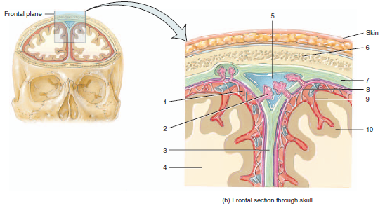
Lab #10 Summer 2020.pdf - ANATOMY AND PHYSIOLOGY I LAB #10... - Course Hero Page 480 Label the meninges and associated structures: Choices to Use: cerebral cortex (cerebrum) periosteal layer of dura mater meningeal layer of dura mater arachnoid mater pia mater subdural space subarachnoid space superior sagittal sinus parietal bone 1. ___PARIETAL BONE_____ 2. ___PERIOSTEAL LAYER_____ 3. Meninges Layers, Function & Anatomy - Study.com These organs are covered by layers of tissues called meninges. There are three layers of meninges: Dura Mater Arachnoid Mater Pia Mater The meninges compose three of the important layers of the... Fundamental the Nervous System an vous Ti - lake.k12.fl.us 1. Figure 12.9 shows a frontal view of the meninges of the brain at the level of the superior sagittal (dural) sinus. First, label arachnoid villi and falx cerebri on the figure. Then, select different colors for each of the following structures and use them to color the diagram. o Dura mater o Pia mater o Arachnoid o Subarachnoid space Scalp Quiz: The Ventricles and Cerebrospinal Fluid - CliffsNotes Quiz: The Ventricles and Cerebrospinal Fluid. Anatomy and Chemistry Basics. Quiz: What is Anatomy and Physiology? Atoms, Molecules, Ions, and Bonds.
Chapter 14 Worksheet Flashcards | Quizlet
Free Science Flashcards about ANP1040 Exam 4 - StudyStack ANP1040 Exam 4. Correctly label the following anatomical features of a neuron. Correctly label the structures, areas, and concentrations associated with a cell's electrical charge difference across its membrane. ___ division carries signals to the smooth muscle in the large intestine.
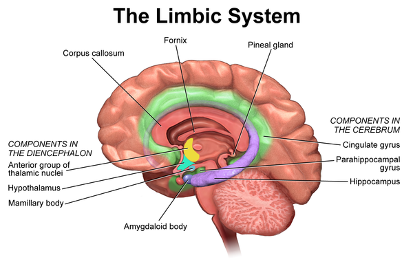
Limbic System - Physiopedia
Ventricles of the Brain: Overview, Gross Anatomy ... - Medscape Overview. The ventricles of the brain are a communicating network of cavities filled with cerebrospinal fluid (CSF) and located within the brain parenchyma. The ventricular system is composed of 2 lateral ventricles, the third ventricle, the cerebral aqueduct, and the fourth ventricle (see the images below). The choroid plexuses are located in ...

Manual sub-segmentation of the cererbellum | medRxiv
Neuroanatomy, Cranial Meninges - StatPearls - NCBI Bookshelf The brain and spinal cord are enveloped within three layers of membrane collectively known as the meninges, with the cranial meninges specifically referring to the section that covers the brain. From superficial to deep, the three layers are the dura, arachnoid, and pia—the term "mater," Latin for mother, often follows these names (i.e., dura mater, arachnoid mater, pia mater).[1] The ...

Meninges and Associated Structures Diagram | Quizlet
Log In to Canvas Forgot Password? Enter your LoLA Username and we'll send you a link to change your password.
PPT - Nervous System PowerPoint Presentation, free download ...
Brain - description - a b c d e f g h i j k l NAME ... - StuDocu Identify the meningeal (or associated) structures described below: 1. outermost meninx covering the brain; composed of tough fibrousconnective tissue 2. innermost meninx covering the brain; delicate and highly vascular 3. structures instrumental in returning cerebrospinal fluid to the venousblood in the dural sinuses 4. structure that forms the ...

Brain injury environment critically influences the ...
Lab 9 Nervous System Anatomy Part 1: Functional Anatomy of the Cerebral ... Write the names of the key anatomical structures of the meninges and ventricles on to the stickers 3. Select a "team leader" and using the colored images as reference have members of the group take turns labeling the sagittal head model with the key anatomical structures. 4. Have your instructor check your work and then move to the next activity.
Solved K The Brain and Cranial Nerves Art-labeling Activity ...
Spinal cord: Anatomy, structure, tracts and function | Kenhub Anatomy. The spinal cord is part of the central nervous system (CNS). It is situated inside the vertebral canal of the vertebral column. During development, there's a disproportion between spinal cord growth and vertebral column growth. The spinal cord finishes growing at the age of 4, while the vertebral column finishes growing at age 14-18.

Techniques for visualizing fibroblast-vessel interactions in ...
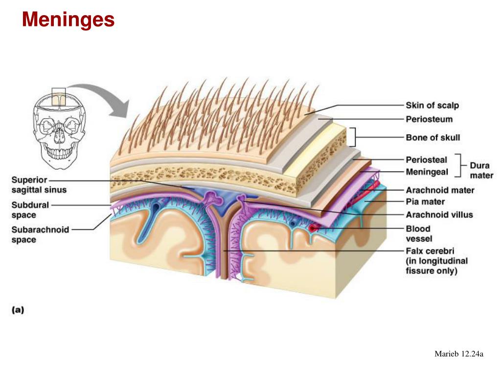
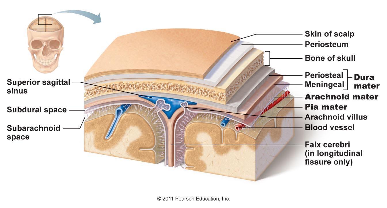
Meninges. Human brain | Human brain, Dura mater, Human anatomy and ... Figure 12.24 Meninges: dura mater, arachnoid mater, and pia mater. Skin of scalp. Periosteum. Bone of skull. Dura mater. Dural venus sinus. Arachnoid mater. Pia mater. Arachnoid villus. Blood vessel. Subarachnoid space. Figure 11.15 The brain. Midbrain. Figure 11.15 The brain. Answers from Science - Neuroscience - Human Brain Anatomy Brain Stem
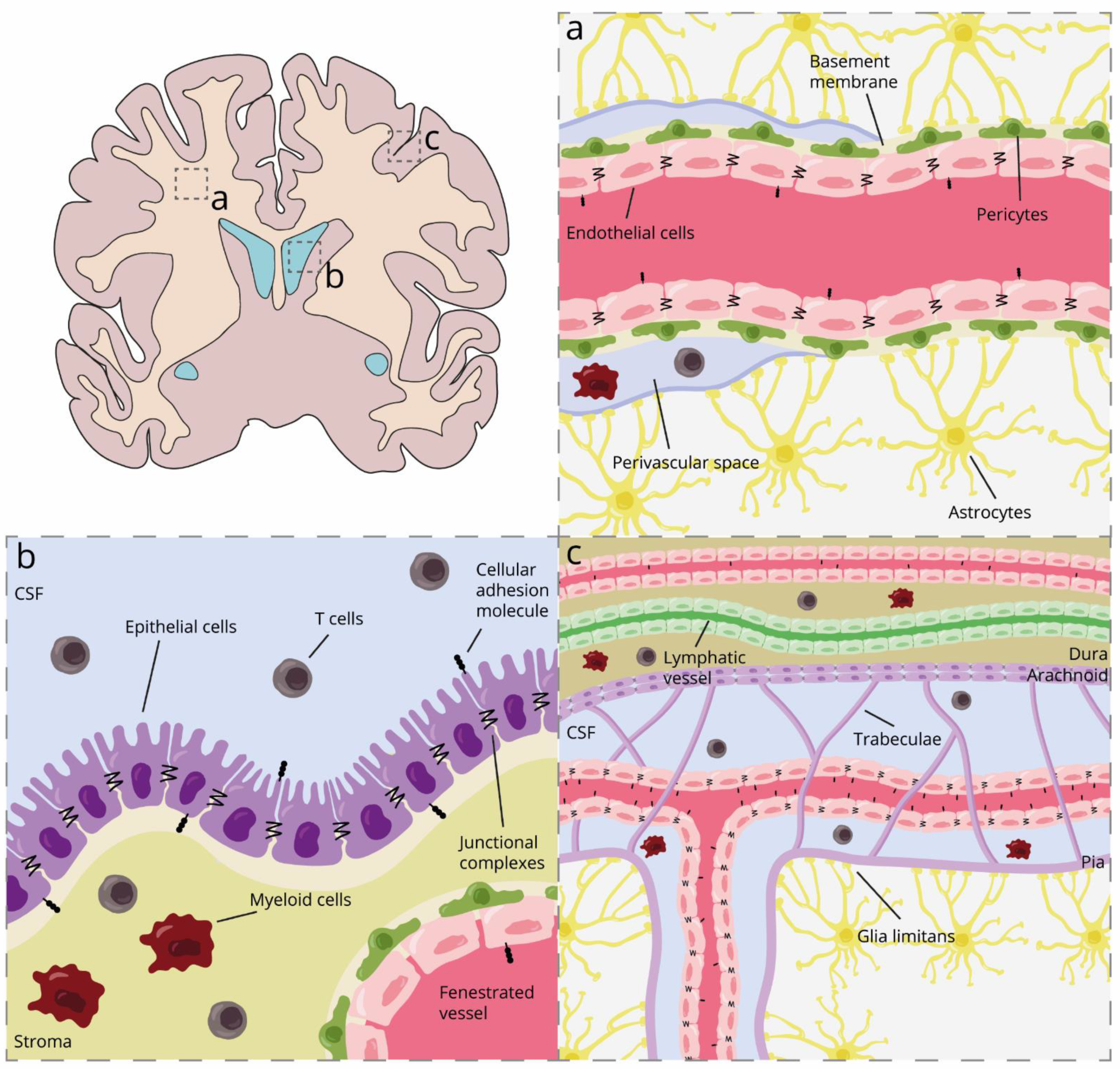
Biomolecules | Free Full-Text | Breaching Brain Barriers: B ...
Meninges: What They Are & Function - Cleveland Clinic Meninges are three membranes layers that cover and protect your brain and spinal cord (central nervous system). These membranes — the dura mater, arachnoid mater and pia mater — protect and anchor your brain and provide a support system for blood vessels, nerves, lymphatics and the cerebrospinal fluid that surrounds your central nervous system.

DB is associated with increased phagocytic activity but not ...
What midbrain structure is a visual reflex center Superior colliculi of the tectal plate The Meninges of the Brain Correctly label the following meninges and associated structures. The Flow of Cerebrospinal Fluid Place a single word into each sentence to make it correct. Not all terms will be used.
quiz 5 part 2 Flashcards | Chegg.com
Label the landmarks of the skull in the figure below Several of these are described on the following pages. Locating Body Landmarks. Anterior Body Landmarks. Identify and use anatomical terms to correctly label the following regions on Figure 1:. i miss you message for her long distance. Bumps and grooves of the brain. In humans, the lobes of the brain are divided by a number of bumps and grooves.

Headache Classification Committee of the International ...
Solved Correctly label the following meninges and associated - Chegg Science. Anatomy and Physiology. Anatomy and Physiology questions and answers. Correctly label the following meninges and associated structures Arachnoid valus Arachnoid maler Meningeal layer of dura Pia mater Subarachnoid space Periosteal layer of dura Fabcccrobni.
Isoniazid | C6H7N3O - PubChem

Drag the correct label to the appropriate location to label ...
Susunan Dewan Redaksi Jurnal Farmasi dan Ilmu ...

Comprehensive characterization of migration profiles of ...
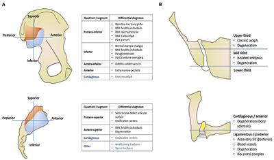
Frontiers | Axial Spondyloarthritis: Mimics and Pitfalls of ...

Chapter 13 Flashcards | Quizlet
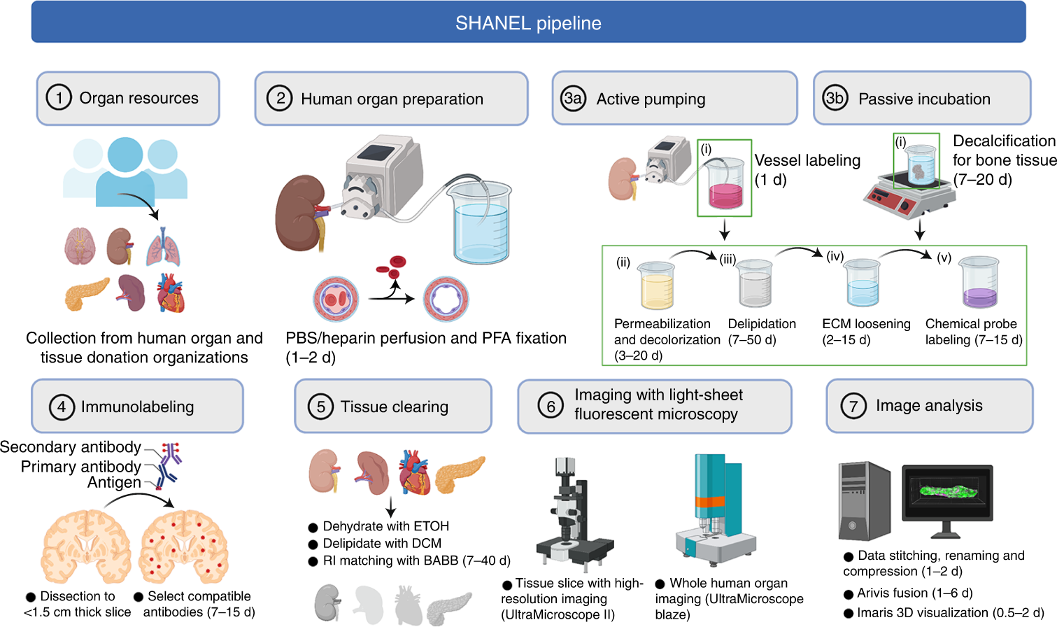
Scalable tissue labeling and clearing of intact human organs ...
Chloramphenicol | C11H12Cl2N2O5 - PubChem
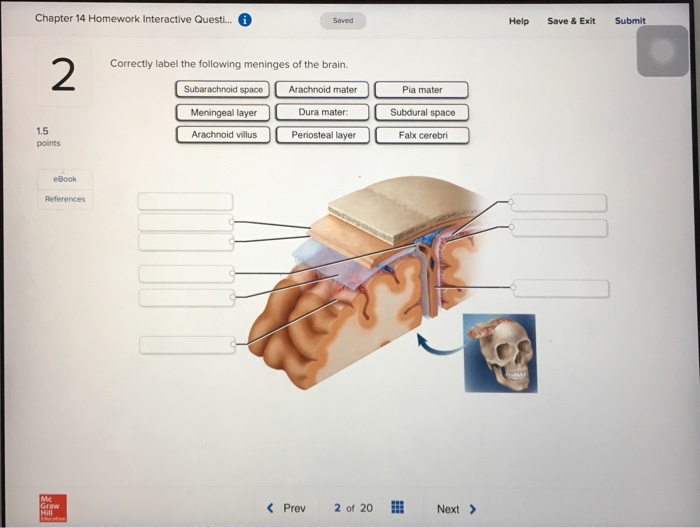
Solved Chapter 14 Homework Interactive Questi.. Help Save ...
Distinct roles of the meningeal layers in CNS autoimmunity ...

Parkinson's Disease-associated α-Synuclein Is a Calmodulin ...

Solved heducation.com Brain and Spinal Cord Correctly label ...

Chapter 14 Worksheet Flashcards | Quizlet
From outermost to innermost, what are the names and the ...

What Is Central Nervous System? Definition, Function & Parts
KUMPULAN ALIH BAHASA DI BIDANG PETERNAKAN DAN KESEHATAN HEWAN ...
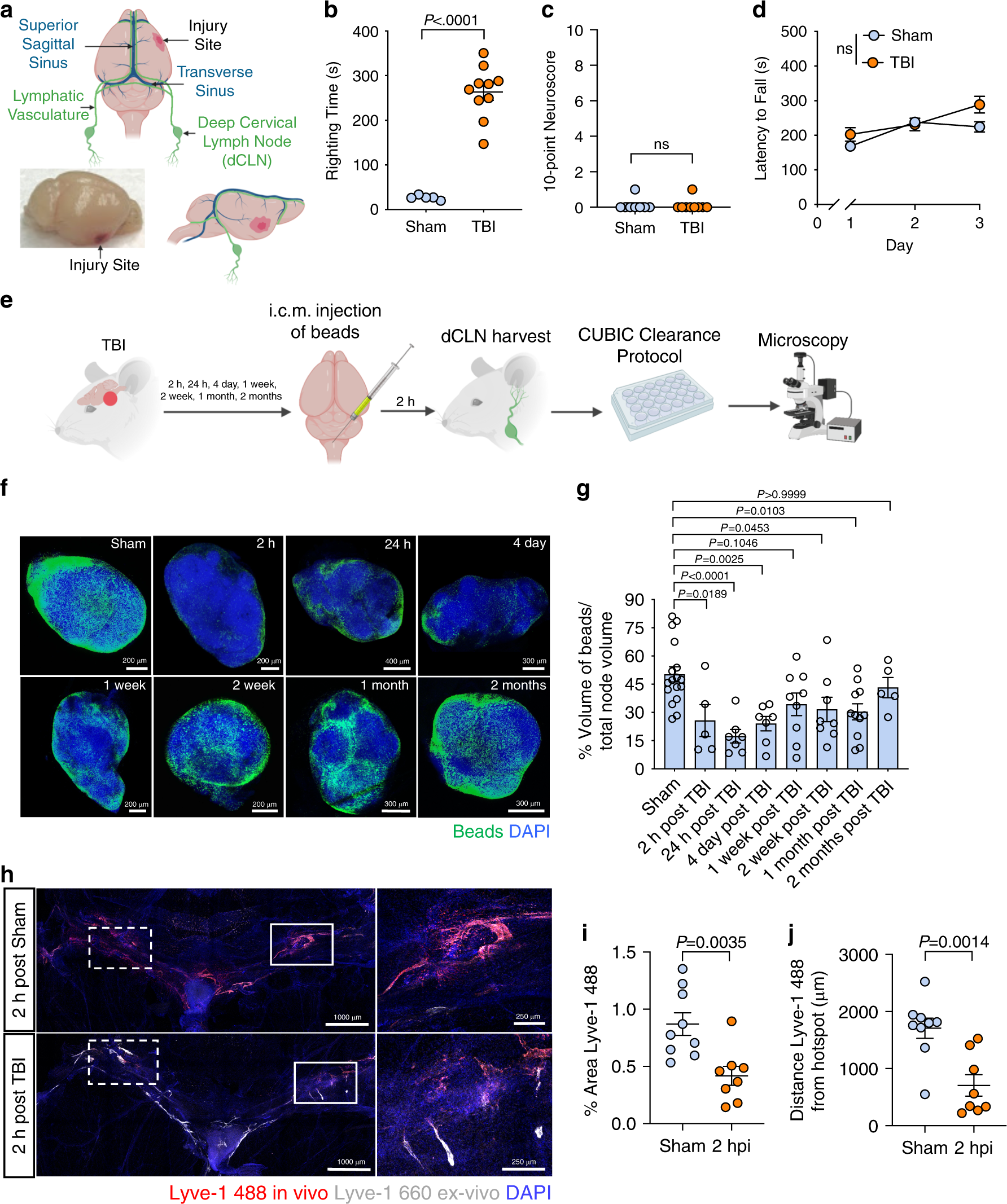
Meningeal lymphatic dysfunction exacerbates traumatic brain ...

Monosialotetrahexosylganglioside Promotes Early Aβ42 Oligomer ...
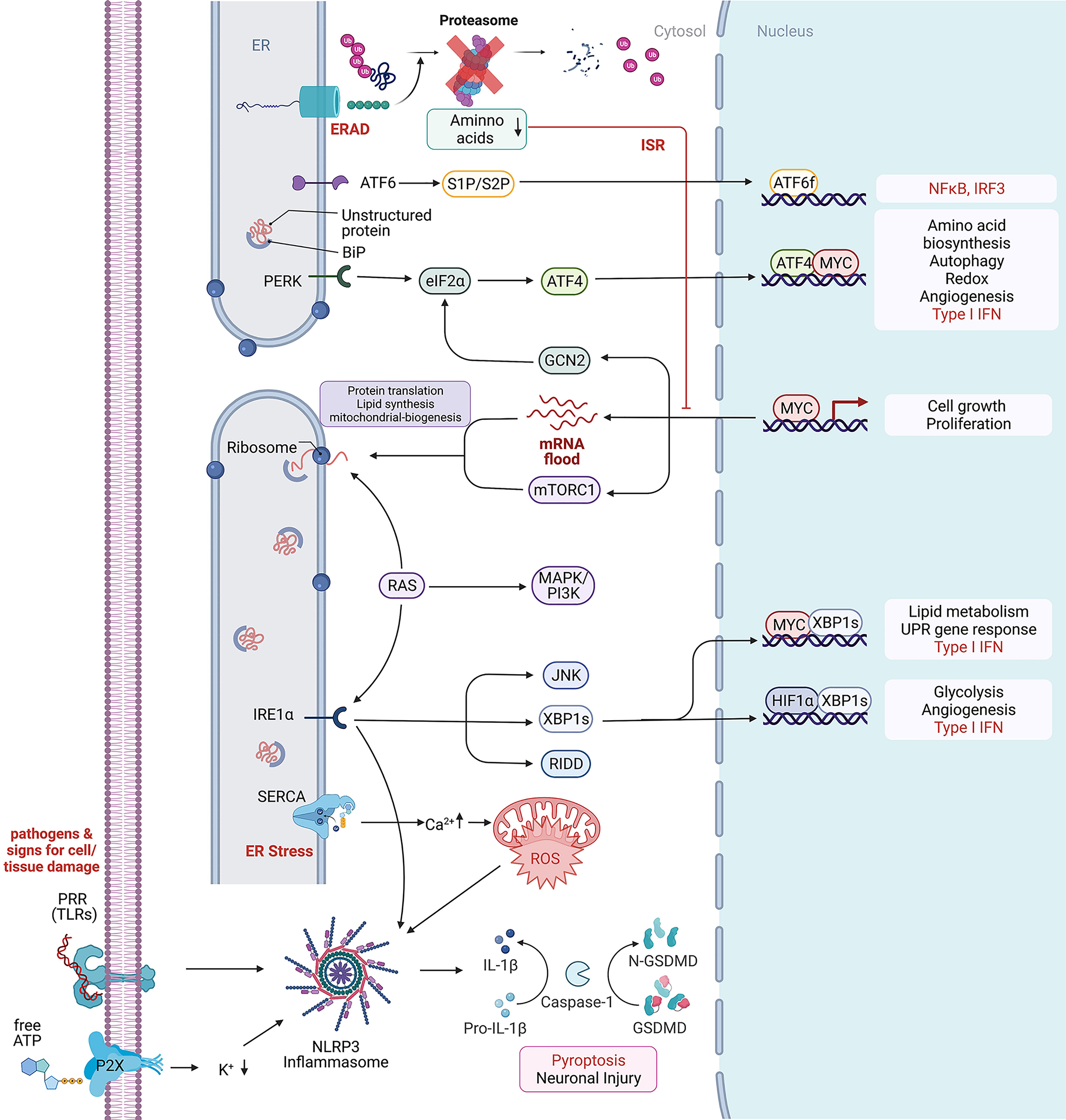
Frontiers | Endoplasmic Reticulum Stress and Its Role in ...
Review of Forensic Medicine and Toxicology, 2nd Edition

Chapter 14 Worksheet Flashcards | Quizlet

Draw the cranial nerves to the brain. Include a chart with ...

A TNF receptor 2 agonist ameliorates neuropathology and ...
Anatomy Exam 2 Flashcards - Easy Notecards

Histology of Central Nervous System Dr. Sama ul Haque. - ppt ...
Leptomeningeal Carcinomatosis

A&P2 FINAL EXAM (1-5) Flashcards | Quizlet
Pengajuan Admin Dapodik Sekolah : SDN DADAP II

Chapter 13 Question Set Flashcards | Quizlet

Correctly Label the Following Meninges and Associated ...
Post a Comment for "42 correctly label the following meninges and associated structures."