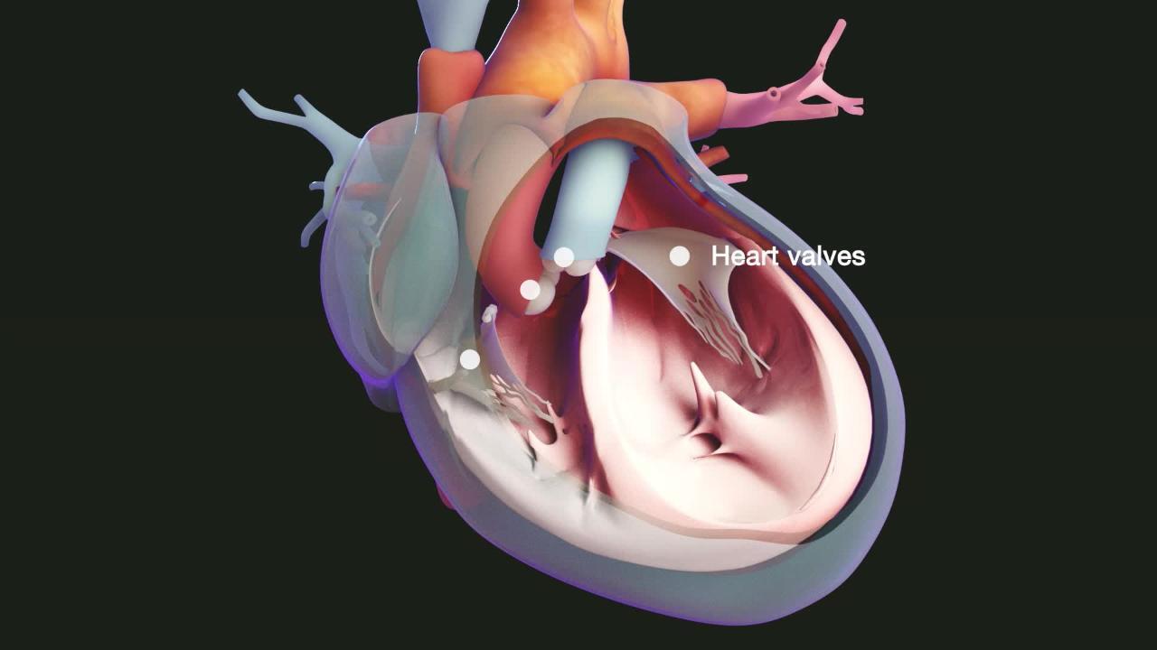
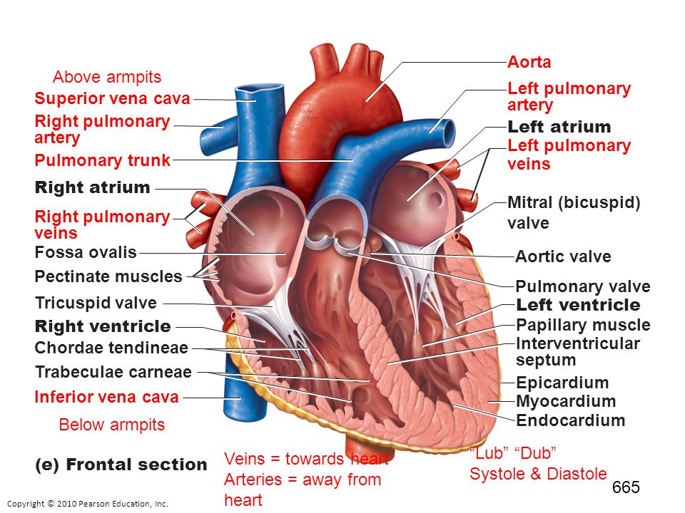
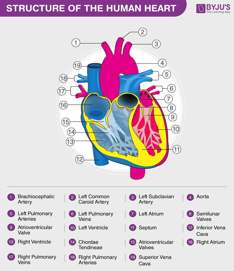
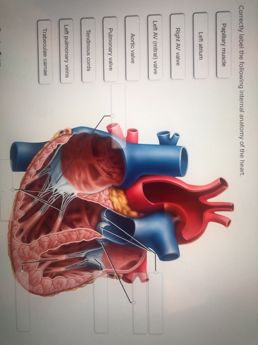
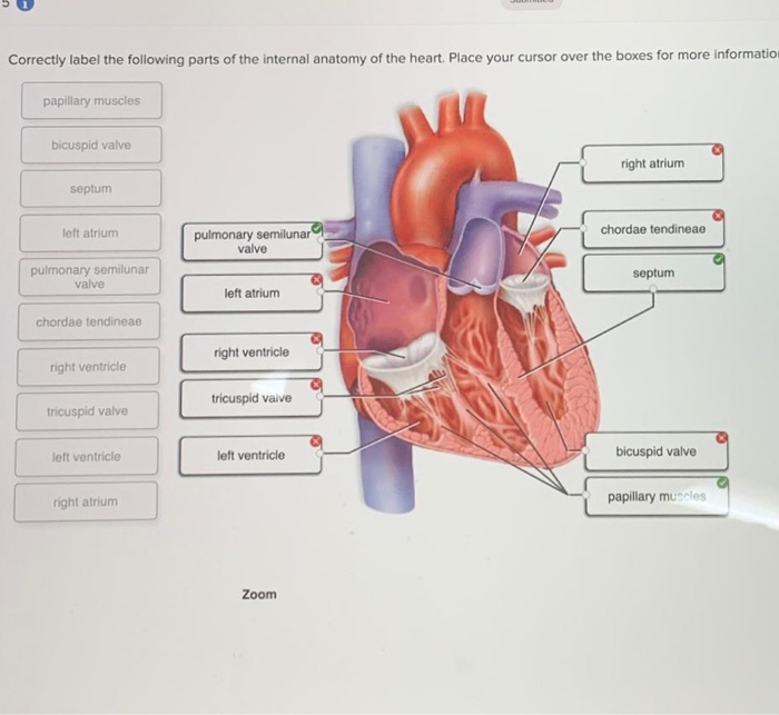
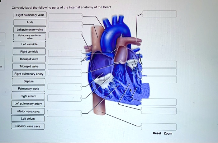
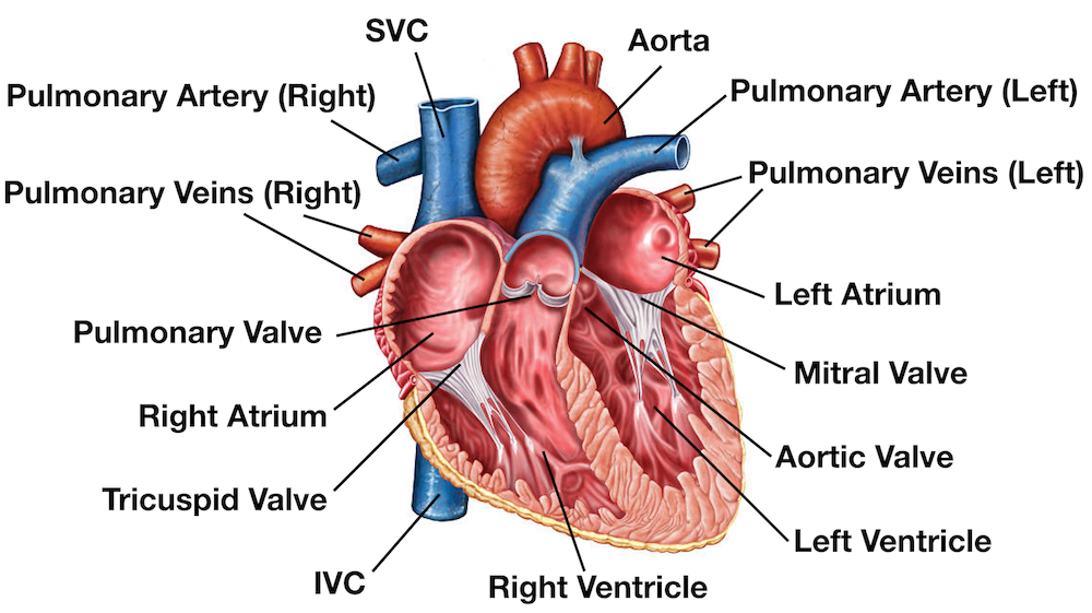
42 correctly label the following internal anatomy of the heart.
Solved Correctly label the following internal anatomy of the ... Correctly label the following internal anatomy of the heart Right atrium Interventricular septum Right ventricle Right AV (tricuspid) valve Tendinous cords Papillary muscles Fossa ovalis Right atrium Pectinate muscles Right AV (tricuspid) valve This problem has been solved! Ch. 19 Circulatory System- heart Flashcards | Quizlet Correctly label the pathway for the cardiac conduction system. Step 1: The sinuatrial (SA) node is a patch of modified cardiomyocytes in the right atrium, just under the epicardium near the superior vena cava. This is the pacemaker that initiates each heartbeat and determines the heart rate.
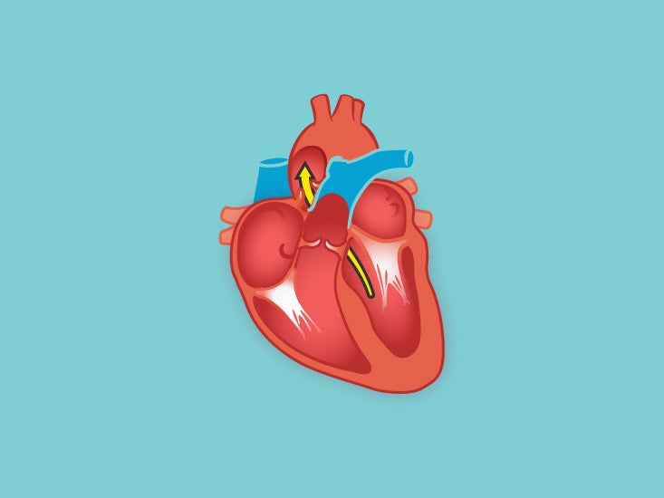
Heart: Anatomy and Function - Cleveland Clinic Your heart walls are the muscles that contract (squeeze) and relax to send blood throughout your body. A layer of muscular tissue called the septum divides your heart walls into the left and right sides. Your heart walls have three layers: Endocardium: Inner layer. Myocardium: Muscular middle layer. Epicardium: Protective outer layer.

Correctly label the following internal anatomy of the heart.
Chapter 20-Cardiovascular System Flashcards | Quizlet Correctly label the following internal anatomy of the heart. b Place the labels in order denoting the flow of oxygenated blood through the heart beginning with the vessels that bring blood back to the heart from the lungs. Correctly label the following coronary blood vessels of the heart. Label Internal Anatomy of The Heart Diagram | Quizlet Start studying Label Internal Anatomy of The Heart. Learn vocabulary, terms, and more with flashcards, games, and other study tools. The Anatomy of the Heart, Its Structures, and Functions Apr 5, 2020 · The heart is made up of four chambers: Atria: Upper two chambers of the heart. Ventricles: Lower two chambers of the heart. Heart Wall The heart wall consists of three layers: Epicardium: The outer layer of the wall of the heart. Myocardium: The muscular middle layer of the wall of the heart. Endocardium: The inner layer of the heart.
Correctly label the following internal anatomy of the heart.. The Anatomy of the Heart, Its Structures, and Functions Apr 5, 2020 · The heart is made up of four chambers: Atria: Upper two chambers of the heart. Ventricles: Lower two chambers of the heart. Heart Wall The heart wall consists of three layers: Epicardium: The outer layer of the wall of the heart. Myocardium: The muscular middle layer of the wall of the heart. Endocardium: The inner layer of the heart. Label Internal Anatomy of The Heart Diagram | Quizlet Start studying Label Internal Anatomy of The Heart. Learn vocabulary, terms, and more with flashcards, games, and other study tools. Chapter 20-Cardiovascular System Flashcards | Quizlet Correctly label the following internal anatomy of the heart. b Place the labels in order denoting the flow of oxygenated blood through the heart beginning with the vessels that bring blood back to the heart from the lungs. Correctly label the following coronary blood vessels of the heart.
:watermark(/images/watermark_5000_10percent.png,0,0,0):watermark(/images/logo_url.png,-10,-10,0):format(jpeg)/images/overview_image/424/Xzp9JjY9J41UO3dz9uTA_structure-of-large-intestine_english.jpg)









:watermark(/images/watermark_only_sm.png,0,0,0):watermark(/images/logo_url_sm.png,-10,-10,0):format(jpeg)/images/anatomy_term/subendocardial-branches-of-atrioventricular-bundle/elMEV9b1krwMpse6Lxkzg_Prukinje_fibers.png)







Post a Comment for "42 correctly label the following internal anatomy of the heart."